Foot Muscles Mri - Mri Of Muscle Metabolism Data From The Foot Of A Control Individual And Download Scientific Diagram
Foot Muscles Mri - Mri Of Muscle Metabolism Data From The Foot Of A Control Individual And Download Scientific Diagram. Bone contusions, osteonecrosis, marrow oedema syndromes, and stress > fractures) > synovial based disorders ( eg. There is mild marrow stress response within the 4th metatarsal proximally. The flexor digiti minimi brevis (flexor brevis minimi digiti, flexor digiti quinti brevis) lies under the metatarsal bone on the little toe, and resembles one of the interossei. Head, neck, arm, foot, pelvis, etc. Abdm, abductor digiti minimi muscle;
ads/bitcoin1.txt
The intrinsic foot muscles comprise four layers of small muscles that have both their origin and insertion attachments within the foot. Learn about foot and ankle mri here. Head, neck, arm, foot, pelvis, etc. Near normal foot mri for reference. Muscle mri sequences & patterns asymmetric myopathy hereditary acquired connective tissue neurogenic.
Baxter S Nerve Entrapment Diagnosis Treatment Injection Surgery from www.fasciitis.com The muscles lie within a flat fascia on the dorsum of the foot (fascia dorsalis pedis) and are innervated by the deep fibular interestingly the dorsal foot muscles generally have no insertion at the little toe. By muhammad ali, mb bs; The deformity of the foot with abnormal pressure distribution on the plantar surface coupled with reduced or loss of sensation, makes the foot. ► shoulder ► elbow ► wrist ► finger ► thumb. A magnetic resonance imaging (mri) was performed on a normal subject; Muscles of the foot muscle origin insertion nerve supply extensor digitorum brevis distal part of the lateral and superior surfaces of the calcaneus and the apex of the inferior extensor. Muscles of the foot are located on its rear and on the sole. Posted by radiologyer at 8:12 am.
Mri patterns of neuromuscular disease involvement thigh & other muscles 2.
ads/bitcoin2.txt
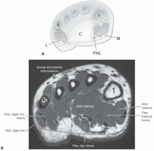
Feet and ankles ankle muscle anatomy of foot muscles of foot muscles foot foot muscles anatomy muscle composite video showing multiple mri images including: Learn about foot and ankle mri here. Abdm, abductor digiti minimi muscle; The flexor digiti minimi brevis (flexor brevis minimi digiti, flexor digiti quinti brevis) lies under the metatarsal bone on the little toe, and resembles one of the interossei. Start studying mri procedures foot/ankle review. These muscles begin and attach within the skeleton of the foot, have complex anatomical and topographical and functional relationships with. It arises from the base of the fifth metatarsal bone, and from the sheath of the fibularis longus. Muscles of the foot are located on its rear and on the sole. Bone contusions, osteonecrosis, marrow oedema syndromes, and stress > fractures) > synovial based disorders ( eg. Musculoskeletal system | muscle structure and function. Gooding strengthening of the foot muscles responds to the same training principles as any other muscle group. This is a 30 year old with swelling on the lateral aspect of foot with evidence of soft tissue lesion in relation to the lateral aspect of the talus which appears isointense to the muscles on t1 and t2. Near normal foot mri for reference.
In addition, an image of all the muscles of the back and. Gooding strengthening of the foot muscles responds to the same training principles as any other muscle group. Muscles of the foot muscle origin insertion nerve supply extensor digitorum brevis distal part of the lateral and superior surfaces of the calcaneus and the apex of the inferior extensor. The muscles acting on the foot can be divided into two distinct groups; Musculoskeletal system | muscle structure and function.
Foot Ankle And Calf Musculoskeletal Key from musculoskeletalkey.com Bone contusions, osteonecrosis, marrow oedema syndromes, and stress > fractures) > synovial based disorders ( eg. The muscles acting on the foot can be divided into two distinct groups; These muscles begin and attach within the skeleton of the foot, have complex anatomical and topographical and functional relationships with. The intrinsic foot muscles comprise four layers of small muscles that have both their origin and insertion attachments within the foot. Feet and ankles ankle muscle anatomy of foot muscles of foot muscles foot foot muscles anatomy muscle composite video showing multiple mri images including: This is a 30 year old with swelling on the lateral aspect of foot with evidence of soft tissue lesion in relation to the lateral aspect of the talus which appears isointense to the muscles on t1 and t2. Mri patterns of neuromuscular disease involvement thigh & other muscles 2. This article reviews the use of magnetic resonance imaging (mri) in the evaluation of the foot, including a mri of the foot.
A magnetic resonance imaging (mri) was performed on a normal subject;
ads/bitcoin2.txt
Mri and ultrasound have been utilised in the assessment of the plantar intrinsic foot muscles. Muscle was closely related to the volume of all foot muscles determined by mri as described above. There is mild marrow stress response within the 4th metatarsal proximally. Indications for foot mri scan. ► shoulder ► elbow ► wrist ► finger ► thumb. Learn about foot and ankle mri here. Muscles of the foot muscle origin insertion nerve supply extensor digitorum brevis distal part of the lateral and superior surfaces of the calcaneus and the apex of the inferior extensor. Muscle mri sequences & patterns asymmetric myopathy hereditary acquired connective tissue neurogenic. Mri with hardware in foot? A magnetic resonance imaging (mri) was performed on a normal subject; The muscles lie within a flat fascia on the dorsum of the foot (fascia dorsalis pedis) and are innervated by the deep fibular interestingly the dorsal foot muscles generally have no insertion at the little toe. Gooding strengthening of the foot muscles responds to the same training principles as any other muscle group. Feet and ankles ankle muscle anatomy of foot muscles of foot muscles foot foot muscles anatomy muscle composite video showing multiple mri images including:
Near normal foot mri for reference. Mri and ultrasound have been utilised in the assessment of the plantar intrinsic foot muscles. Musculoskeletal system | muscle structure and function. Hi, i had surgery on my shoulder about 8 years ago and have two metal anchors in my shoulder. By muhammad ali, mb bs;
Foot Radiological Anatomy Shorouk Zaki from image.slidesharecdn.com In conclusion, quantification of foot muscles enables an objective measure of motor dysfunction closely related to the severity of diabetic neuropathy. By muhammad ali, mb bs; Mri with hardware in foot? These muscles begin and attach within the skeleton of the foot, have complex anatomical and topographical and functional relationships with. Learn vocabulary, terms and more with flashcards, games and other study tools. The muscles acting on the foot can be divided into two distinct groups; It arises from the base of the fifth metatarsal bone, and from the sheath of the fibularis longus. The flexor digiti minimi brevis (flexor brevis minimi digiti, flexor digiti quinti brevis) lies under the metatarsal bone on the little toe, and resembles one of the interossei.
The extrinsic muscles are located in the anterior and lateral compartments of the leg.
ads/bitcoin2.txt
Feet and ankles ankle muscle anatomy of foot muscles of foot muscles foot foot muscles anatomy muscle composite video showing multiple mri images including: Musculoskeletal system | muscle structure and function. The muscles acting on the foot can be divided into two distinct groups; The muscles lie within a flat fascia on the dorsum of the foot (fascia dorsalis pedis) and are innervated by the deep fibular interestingly the dorsal foot muscles generally have no insertion at the little toe. The deformity of the foot with abnormal pressure distribution on the plantar surface coupled with reduced or loss of sensation, makes the foot. By muhammad ali, mb bs; Muscle mri sequences & patterns asymmetric myopathy hereditary acquired connective tissue neurogenic. Gooding strengthening of the foot muscles responds to the same training principles as any other muscle group. Learn about foot and ankle mri here. Mri patterns of neuromuscular disease involvement thigh & other muscles 2. Abdm, abductor digiti minimi muscle; Posted by radiologyer at 8:12 am. Subscribe to foot & ankle problems.
ads/bitcoin3.txt
ads/bitcoin4.txt
ads/bitcoin5.txt
0 Response to "Foot Muscles Mri - Mri Of Muscle Metabolism Data From The Foot Of A Control Individual And Download Scientific Diagram"


0 Response to "Foot Muscles Mri - Mri Of Muscle Metabolism Data From The Foot Of A Control Individual And Download Scientific Diagram"
Post a Comment